


Oral cancer screening is a clinical examination designed to detect early signs of mouth cancer and precancerous changes before symptoms become advanced. During the exam, a dental professional visually inspects the lips, tongue, cheeks, floor of the mouth, palate, and throat, and may palpate tissues to identify abnormal lumps or lesions. Screening is quick, painless, and typically performed during routine dental visits. Early detection significantly improves treatment outcomes, as many oral cancers are asymptomatic in their initial stages. Individuals who use tobacco, consume alcohol regularly, or have HPV exposure may require closer monitoring.
Oral cancer can develop silently. In its earliest stages, it may not cause pain, noticeable swelling, or difficulty eating. Because early lesions are often subtle, routine oral cancer screening plays a critical role in early detection.
A screening exam does not diagnose cancer directly. Instead, it identifies suspicious abnormalities that may require further evaluation.
At Smiles By Design Dentistry of San Diego, comprehensive dental examinations routinely include visual and tactile screening of oral tissues. Early identification can significantly influence prognosis.
Oral cancer screening is a preventive clinical examination aimed at identifying abnormal tissue changes in the mouth and throat that may indicate precancerous or cancerous conditions.
The goal is early detection before symptoms become advanced.
Screening does not replace biopsy or imaging but serves as an initial assessment tool.
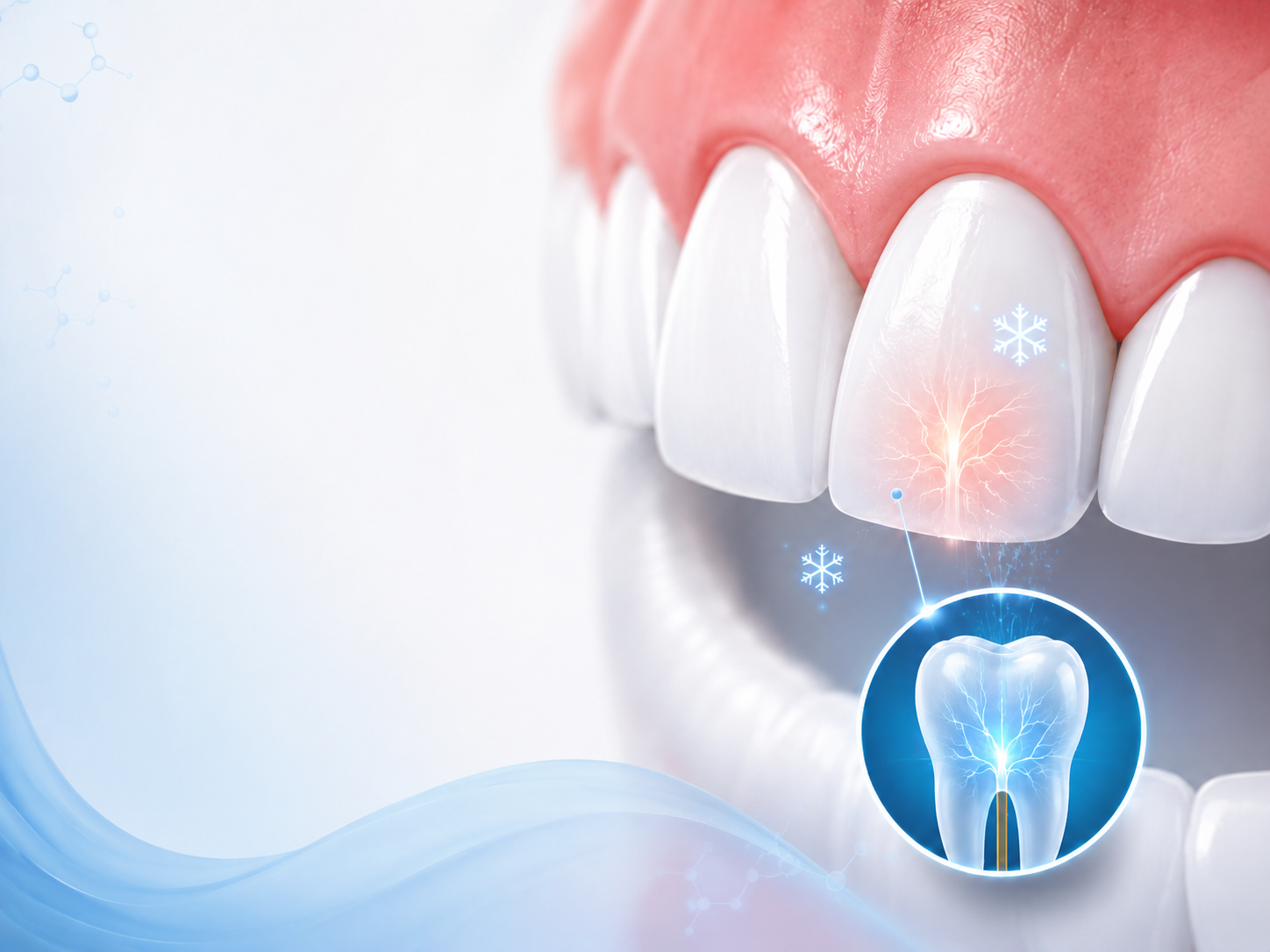
Oral cancer most commonly arises from squamous cells lining the mouth and throat. Chronic exposure to carcinogens—such as tobacco smoke or excessive alcohol—can cause cellular mutations.
These mutations may lead to dysplasia (precancerous cellular changes) and, over time, invasive carcinoma.
Because cellular transformation begins microscopically, visible lesions may appear only after significant tissue alteration.
Screening seeks to detect surface abnormalities, color changes, texture differences, and tissue firmness that could reflect underlying cellular transformation.
Risk factors include:
However, oral cancer can occur in individuals without these risk factors.
Risk assessment influences screening frequency.
Possible warning signs include:
Many early lesions are painless.
Any abnormality lasting longer than two weeks warrants evaluation.
The mouth naturally contains variations in pigmentation, minor ridges, and harmless tissue irregularities.
Benign conditions such as geographic tongue or minor aphthous ulcers are common and typically self-limiting.
Suspicious lesions often display:
Distinguishing normal variation from pathological change requires professional assessment.
Evaluation is recommended if:
Early consultation improves diagnostic timelines.
Screening includes:
If abnormalities are detected, further diagnostic steps may include biopsy or referral to a specialist.
The exam is typically performed during a routine dental visit.
The clinician uses a light source and gauze to move the tongue and examine all surfaces. Gloves are worn to gently palpate tissues for firmness or irregularity.
Some offices may use adjunctive screening devices, such as special lights or dyes, though visual and tactile examination remains the primary method.
The entire process usually takes only a few minutes.
Adjunct tools may include:
These tools assist in highlighting suspicious areas but do not replace conventional biopsy for definitive diagnosis.
Clinical judgment remains central.
Oral cancer screening is non-invasive and carries minimal risk.
However, limitations include:
Screening improves early detection but does not guarantee identification of all cancers.
Regular exams enhance accuracy over time.
When detected early, oral cancer has significantly higher survival rates compared to late-stage diagnosis.
Advanced oral cancers may require extensive surgery, radiation, or chemotherapy.
Early-stage lesions often allow more conservative treatment.
Timely screening therefore directly influences treatment complexity and survival outcomes.
Preventive strategies include:
While not all oral cancers are preventable, risk reduction significantly lowers incidence.
Oral cancer screening is not painful.
It is not only for high-risk individuals.
It does not automatically mean cancer is suspected.
A lack of pain does not rule out serious disease.
Education reduces fear-based avoidance.
How often should I get an oral cancer screening?
Most adults receive screening during routine dental visits, typically every six months.
Is oral cancer screening painful?
No, it is a visual and tactile examination.
Can oral cancer be detected early?
Yes, many early lesions are visible before symptoms develop.
Do I need screening if I don’t smoke?
Yes, because oral cancer can occur in non-smokers as well.
Dr. Dan Javaheri, a graduate of the New York University College of Dentistry, emphasizes that routine oral cancer screening should be integrated into comprehensive dental examinations rather than performed only when symptoms arise. With ongoing education in diagnostic and restorative care and certification through the California Academy of Implant Dentistry, he prioritizes early lesion detection through systematic visual and tactile assessment. Research involvement associated with the National Institutes of Health and UC Davis Medical Center supports an evidence-based understanding of inflammatory and cellular changes. At Smiles By Design Dentistry of San Diego, preventive screening is incorporated routinely to support early identification and improved patient outcomes.